The term onychomycosis (toenail and toenail fungus) describes a fungal infection of the nails caused by dermatophytes, non-dermatophyte molds or yeasts. There are four clinically distinct forms of onychomycosis. Diagnosis is based on examination with CON, microscopy and histology. Often, treatment includes systemic and local therapy, sometimes using surgical removal.

Factors that contribute to nail fungus
- Increased sweating (hyperhidrosis).
- Vascular deficiency. Violation of the structure and tone of the veins, especially the veins of the lower leg (typical toenail onychomycosis).
- Age. The incidence of the disease in humans increases with age. In 15-20% of the population, pathology occurs at the age of 40-60 years.
- Diseases of internal organs. Disorders of the nervous system, endocrine (most often onychomycosis occurs in people with diabetes) or the immune system (immunosuppression, especially HIV infection).
- A large nail mass, consisting of a thick nail plate and the contents underneath, can cause discomfort when wearing shoes.
- Traumatization. Continuous trauma to the nail or injury and lack of proper treatment.
Disease prevalence
Onychomycosis– the most common nail disease, which is the cause of 50% of all cases of onychodystrophy (destruction of the nail plate). It affects up to 14% of the population, and both the prevalence of the disease in older people and the overall incidence are increasing. The incidence of onychomycosis in children and adolescents is also increasing; Onychomycosis accounts for 20% of dermatophyte infections in children.
The increased prevalence of this disease may be associated with the wearing of tight shoes, the increased number of people taking immunosuppressive therapy, and the increased use of public locker rooms.
Nail disease usually begins with tinea pedis before spreading to the base of the nail, where eradication is difficult. This area serves as a reservoir for local relapse or spread of infection to other areas. Up to 40% of patients with onychomycosis of the toes have combined skin infections, most often tinea pedis (about 30%).
The causative agent of onychomycosis
In most cases, onychomycosis is caused by dermatophytes, with T. rubrum and T. interdigitale being the causative agents of infection in 90% of all cases. T. tonsurans and E. floccosum have also been documented as etiologic agents.
Yeasts and non-dermatophyte mold organisms such as Acremonium, Aspergillus, Fusarium, Scopulariopsis brevicaulis and Scytalidium are the cause of toe onychomycosis in about 10% of cases. It is interesting to note that Candida species are the causative agent in 30% of cases of finger onychomycosis, while non-dermatophytic molds are not present in affected nails.
Pathogenesis
Dermatophytes have a wide variety of enzymes that, acting as virulence factors, ensure pathogen attachment to the nail. The first stage of infection is attachment to keratin. Due to the further breakdown of keratin and the release of cascade mediators, an inflammatory response develops.

The stages of fungal infection pathogenesis are as follows.
Adhesion
The fungus overcomes several lines of host defense before hyphae begin to live in keratinized tissue. The first is the successful adhesion of arthroconidia to the keratin tissue surface. Early nonspecific lines of host defense include fatty acids in sebum, as well as competitive bacterial colonization.
Several recent studies have examined the molecular mechanisms involved in the adhesion of arthroconidia to keratin surfaces. Dermatophytes have been shown to selectively use their proteolytic reserves during adhesion and invasion. Some time after adhesion has occurred, spores germinate and move to the next stage - invasion.
Invasion
Traumatization and maceration are good environments for fungal penetration. The invasion of elements of fungal growth ends with the release of various proteases and lipases, in general, various products that serve as nutrients for fungi.
The owner's reaction
Fungi face various protective barriers in the host, such as inflammatory mediators, fatty acids, and cellular immunity. The first and most important barrier is the keratinocyte, which is encountered by the invading fungal element. The role of keratinocytes: proliferation (to increase desquamation of horny scales), secretion of antimicrobial peptides, anti-inflammatory cytokines. Once the fungus penetrates deeper, more and more new non-specific mechanisms are activated for protection.
The severity of the host's inflammatory response depends on the immune status, as well as on the natural habitat of the dermatophytes involved in the invasion. The next level of defense is the delayed-type hypersensitivity reaction, which is caused by cell-mediated immunity.
The inflammatory response associated with this hypersensitivity is associated with clinical destruction, while defects in cell-mediated immunity can lead to chronic and recurrent fungal infections.
Although epidemiological observations indicate a genetic predisposition to fungal infections, no molecularly proven studies exist.
Clinical picture and symptoms of damage to toenails and fingernails
There are four characteristic clinical forms of the infection. This form can be isolated or include several clinical forms.
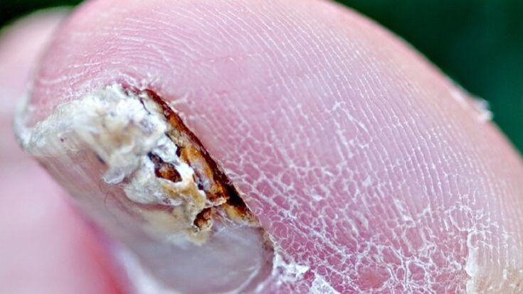
Distal-lateral subungual onychomycosis
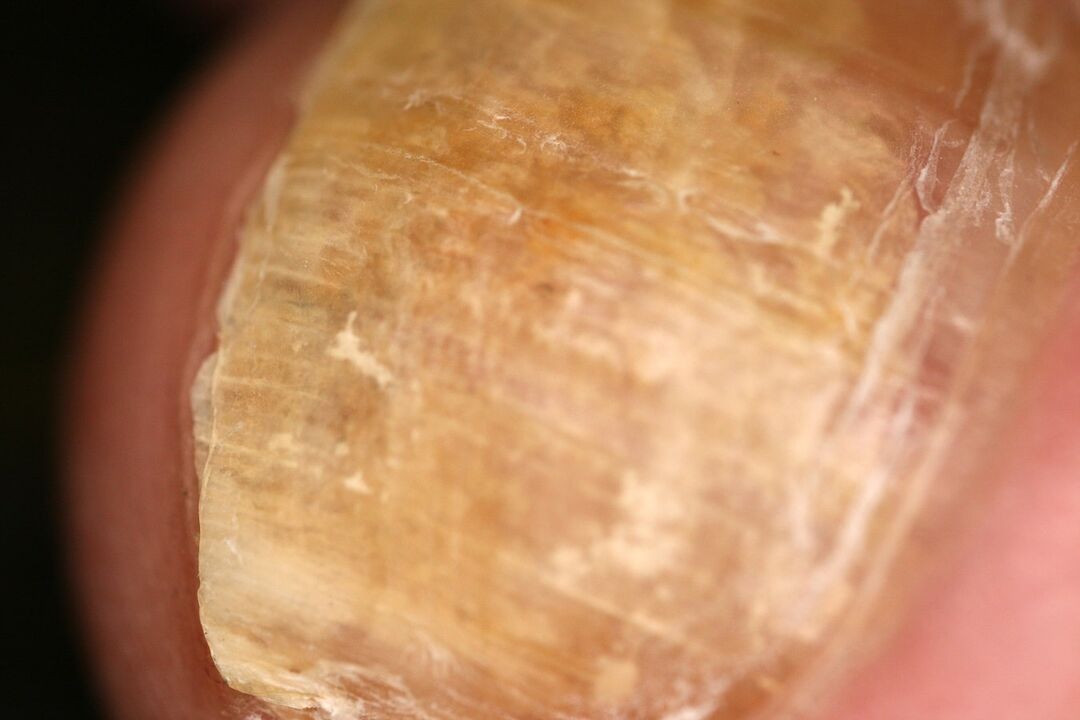
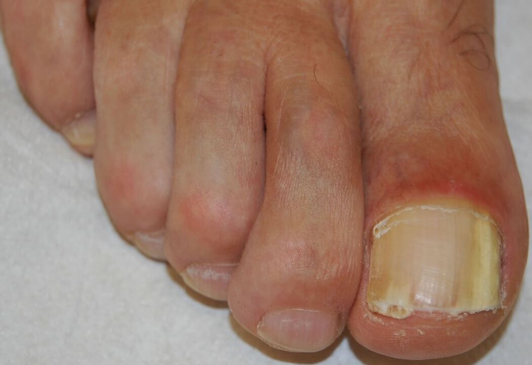
It is the most common form of onychomycosis and can be caused by any of the pathogens listed above. It begins with invasion of the pathogen in the hyponychium stratum corneum and the base of the distal nail, resulting in a whitish or brownish-yellow opacity at the distal end of the nail. The infection then spreads proximally over the base of the nail to the ventral aspect of the nail plate.

Hyperproliferation or impaired differentiation in the nail bed in response to infection causes subungual hyperkeratosis, while progressive invasion of the nail plate leads to increased nail dystrophy.
Proximal subungual onychomycosis
It occurs as a result of infection of the proximal nail fold, mainly by the organisms T. rubrum and T. megninii. Clinic: the proximal part of the nail is cloudy with a white or beige color. This opacity gradually increases and involves the entire nail, eventually leading to leukonychia, proximal onycholysis and/or destruction of the entire nail.
Patients with proximal subungual onychomycosis should be examined for HIV infection, as this form is considered a marker of this disease.
Superficial white onychomycosis
It occurs due to direct invasion of the dorsal nail plate and appears as a dull white or yellow spot, evident on the surface of the toenail. The pathogens are usually T. interdigitale and T. mentargophytes, although non-dermatophyte molds such as Aspergillus, Fusarium and Scopulariopsis are also known to be pathogenic in this form. Candida species can invade the hyponychium epithelium and eventually infect the nail throughout the entire thickness of the nail plate.
Candidal onychomycosis
Damage to the nail plate caused by Candida albicans is observed exclusively in chronic mucocutaneous candidiasis (a rare disease). Usually all nails are affected. The nail plate thickens and acquires various shades of yellow-brown color.
Diagnosis of onychomycosis
Although onychomycosis accounts for 50% of cases of nail dystrophy, it is advisable to obtain laboratory confirmation of the diagnosis before starting toxic systemic antifungal drugs.
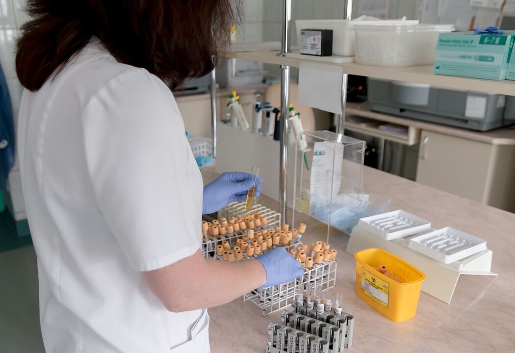
Study of subungual masses with KOH, analysis of nail plate material culture and subungual masses on Sabouraud dextrose agar (with and without antimicrobial additives) and staining of nail sections using the PAS method are the most informative methods.
Learn with CON
It is a standard test for suspected onychomycosis. However, it often gives negative results even with a high index of clinical suspicion, and culture analysis of nail material in which hyphae are found during studies with CON is often negative.
The most reliable way to minimize false negative results due to sampling error is to increase the sample size and resampling.
Cultural analysis
This laboratory test determines the type of fungus and determines the presence of dermatophytes (organisms that respond to antifungal drugs).

To distinguish pathogens from contaminants, the following recommendations are offered:
- if a dermatophyte is isolated in culture, it is considered a pathogen;
- Nondermatophytic molds or yeast organisms isolated in culture are relevant only if hyphae, spores, or yeast cells are observed under the microscope and repeated active growth of nondermatophytic mold pathogens is observed without isolation.
Culture analysis, PAS - the method of staining nail clippings is the most sensitive and does not require waiting for results for several weeks.
Pathological examination
During pathological examination, hyphae are located between the layers of the nail plate, parallel to the surface. In the epidermis, spongiosis and focal parakeratosis, as well as inflammatory reactions, can be observed.
In superficial white onychomycosis, microorganisms are found superficially behind the nail, displaying their unique "perforate organ" pattern and modified hyphal elements called "bitten leaves. "With candidal onychomycosis, invasion of pseudohyphae is observed. Histological examination of onychomycosis occurs with special dyes.
Differential diagnosis of onychomycosis
| Most probably | Sometimes it is possible | Rarely found |
|---|---|---|
|
|
Melanoma |
Treatment methods for nail fungus
Treatment for nail fungus depends on the severity of the nail lesion, the presence of associated tinea pedis, and the effectiveness and potential side effects of the treatment regimen. If nail involvement is minimal, local therapy is a rational decision. When combined with foot dermatophytosis, especially against the background of diabetes mellitus, it is important to prescribe therapy.
Topical antifungal medication
In patients with distal nail involvement or contraindications for systemic therapy, local therapy is recommended. However, we must remember that only local therapy with antifungal agents is not effective enough.
Varnish from the oxypyridone group is gaining popularity, which is used daily for 49 weeks, mycological cure is achieved in about 40% of patients, and nail cleaning (clinical cure) in 5% of cases of mild or moderate onychomycosis caused by dermatophytes.
Although the effectiveness is much lower compared to systemic antifungal drugs, the local use of the drug avoids the risk of drug-drug interactions.
Another drug, specially developed in the form of nail polish, is used 2 times a week. It is a representative of a new class of antifungal drugs, morpholine derivatives, active against yeasts, dermatophytes and molds that cause onychomycosis.
This product may have a higher mycological cure rate than previous varnishes; however, controlled studies are needed to determine statistically significant differences.
Antifungal drugs for oral administration
Systemic antifungal medication is required in cases of onychomycosis involving areas of the matrix, or if a shorter course of treatment or a higher chance of clearance and healing is desired. When choosing an antifungal drug, one should first take into account the etiology of the pathogen, potential side effects and the risk of drug interactions in each individual patient.
Drugs from the allylamine group, which have a fungistatic and fungicidal effect against dermatophytes, Aspergillus, are less effective against Scopulariopsis. This product is not recommended for candidal onychomycosis because it shows variable efficacy against Candida species.
A standard dose of 6 weeks is effective for most toenail injections, while a minimum of 12 weeks is required for toenail injections. Most side effects are related to digestive system problems, including diarrhea, nausea, taste changes, and increased liver enzymes.
Data indicate that a 3-month continuous dosing regimen is currently the most effective systemic therapy for toenail onychomycosis. Clinical cure rates in various studies are approximately 50%, although treatment rates are higher in patients over 65 years of age.
Drugs from the azole group that have a fungistatic effect on dermatophytes, as well as non-dermatophyte molds and yeast organisms. Safe and effective regimens include daily pulse dosing for one week for a month or continuous daily dosing, both of which require two months or two cycles of therapy for the nail and at least three months or three pulses. therapy for toenail lesions.
In children, this medicine is given individually depending on body weight. Although the drug has a wider spectrum of action than its predecessor, studies have shown a much lower cure rate with it and a higher relapse rate.
Elevated liver enzyme levels occur in less than 0. 5% of patients during therapy and return to normal within 12 weeks after discontinuation of treatment.
Drugs that act fungistatically against dermatophytes, some non-dermatophyte molds and Candida species. This medicine is usually taken once a week for 3 to 12 months.
There are no clear criteria for laboratory monitoring of patients receiving the above drugs. It makes sense to do a complete blood count and liver function tests before treatment and 6 weeks after starting treatment.
Medicines from the grisan group are no longer considered standard therapy for onychomycosis due to long treatment, potential side effects, drug interactions and relatively low cure rates.
Combination therapy regimens may result in higher clearance rates than systemic or topical therapy alone. Taking allylamine drugs in combination with the use of morpholine varnish resulted in clinical cure and negative mycological test results in about 60% of patients, compared to 45% of patients who only received systemic allylamine antifungal drugs. However, other studies have shown no additional benefit when combining systemic allylamine agents with oxypyridone drug solutions.
Other medicines
The in vitro demonstrated fungicidal activity of thymol, camphor, menthol and Eucalyptus citriodora oil suggests potential additional therapeutic strategies in the treatment of onychomycosis. Thymol alcohol solution can be used in the form of drops on the nail plate and hyponychia. The use of local preparations with thymol for nails leads to healing in isolated cases.
Surgery
Final treatment options for treatment-resistant cases include surgical removal of the nail with urea. To remove more of the crumbling mass from the affected nail, special clamps are used.
Many doctors believe that the main and first method of treating nail fungus is the mechanical removal of the nail. Often, surgical removal of the affected nail is recommended, and less often, removal using keratolytic patches.
Traditional methods in the fight against nail fungus
Although there are many different folk recipes to get rid of nail fungus, dermatologists do not recommend choosing this treatment option and start with "home diagnosis. "It is wiser to start therapy with local drugs that have undergone clinical trials and proven effective.
Course and prognosis
Bad prognostic signs are pain that appears due to thickening of the nail plate, the addition of secondary bacterial infections, and diabetes mellitus. The most beneficial way to reduce the likelihood of relapse is to combine treatment methods. Therapy for onychomycosis is a long road that does not always lead to complete recovery. However, do not forget that the effect of systemic therapy is up to 80%.
Prevention
Prevention includessome events, thanks to which you can significantly reduce the percentage of onychomycosis infections and reduce the likelihood of relapse.
- Disinfection of personal and public items.
- Systematic disinfection of shoes.
- Treatment of feet, hands, folds (under favorable conditions - favorite localization) with local antifungal agents with the recommendation of a dermatologist.
- If the diagnosis of onychomycosis is confirmed, it is necessary to visit the doctor for monitoring every 6 weeks and after the completion of systemic therapy.
- If possible, at each visit to the doctor you should clean the nail plate.
Conclusion
Onychomycosis (nail and toenail fungus) is an infection caused by various fungi. This disease affects the nail plate of the finger or toe. When making a diagnosis, examine all the skin and nails, and also exclude other diseases that mimic onychomycosis. If there is any doubt about the diagnosis, it must be confirmed either by culture (preferred) or by histological examination of nail sections followed by staining.
Therapy includes surgical removal, local and general medications. Treatment of onychomycosis is a long process that can last for several years, so you cannot expect recovery "from one pill. "If you suspect nail fungus, consult a specialist to confirm the diagnosis and set an individual treatment plan.


